
What is the tibialis posterior tendon?
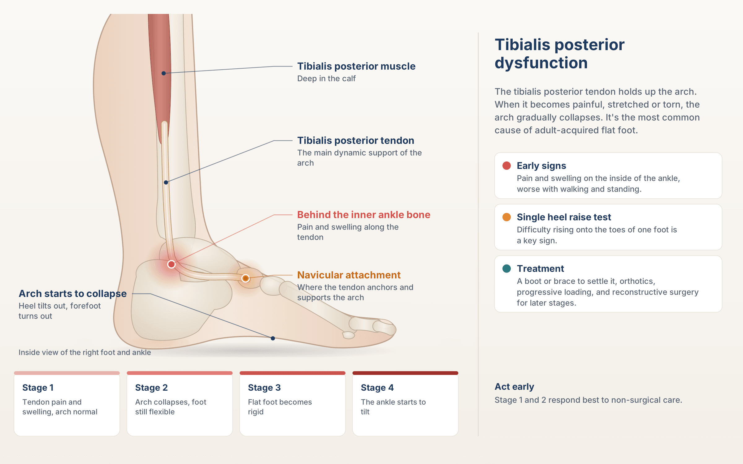
The tibialis posterior muscle sits deep in the calf. Its tendon runs down behind the bony bump on the inside of the ankle (the medial malleolus) and attaches to the navicular bone and other bones in the middle of the foot. It holds up the arch, stabilises the heel, and lets you rise onto your toes.
Tibialis posterior tendon dysfunction (PTTD) happens when the tendon becomes inflamed, degenerates, stretches or tears, so it can no longer support the arch properly. It’s now also called progressive collapsing foot deformity.
Stages of tibialis posterior dysfunction
- Stage 1: pain and swelling along the tendon, with the arch still normal and the foot flexible
- Stage 2: the arch collapses and the heel tilts outward, but the foot is still flexible
- Stage 3: the flat foot becomes rigid, with arthritis in the joints below the ankle
- Stage 4: the ankle joint itself starts to tilt and wear
The earlier it’s treated, the better the chance of stopping it progressing.
What causes tibialis posterior dysfunction?
- Overuse and degeneration of the tendon, most commonly in people over 40
- Pre-existing flat feet, which strain the tendon
- Being overweight
- Diabetes and high blood pressure
- Inflammatory arthritis
- Steroid medication or injections near the tendon
- Injury, such as an ankle sprain or a fall
- Being female, with the condition more common in middle-aged women
- An accessory navicular bone, which can weaken the tendon’s attachment
Symptoms to look out for
- Pain and swelling along the inside of the ankle and arch
- An arch that’s getting flatter, often in one foot
- The heel tilting outward, and the toes pointing outward when viewed from behind (“too many toes” sign)
- Difficulty or pain rising onto your toes on one leg
- Pain when walking, especially on uneven ground, or standing for long periods
- In later stages, pain on the outside of the ankle, where the heel bone presses against the ankle bone
How is it diagnosed?
Your podiatrist will examine the tendon for tenderness, swelling and thickening, look at your feet from behind while you stand, and ask you to perform a single-leg heel raise, one of the most important tests for tendon function. We assess whether the foot is still flexible, check calf flexibility and look at how you walk. Ultrasound or MRI shows the condition of the tendon and any tears, and weight-bearing X-rays assess the alignment of the foot and any arthritis.
Stage 1 to early stage 2: protect and strengthen
- Immobilisation, for acute pain, with a short period in a walking boot to let the tendon settle
- Bracing: an ankle brace or custom ankle-foot orthosis to support the tendon and arch
- Custom orthotics, made from a 3D scan of your feet, with firm arch and heel support
- A progressive loading program for the tibialis posterior, foot and calf muscles, which is a key part of treatment
- Calf stretching where the calf is tight
- Footwear: supportive shoes with a firm heel counter
- Shockwave therapy for long-standing tendon pain, usually over a course of 3 to 5 weekly sessions
- Weight management and activity changes where relevant
We avoid corticosteroid injections into or around the tibialis posterior tendon, as they can weaken it and increase the risk of rupture.
When surgery is needed
If the tendon doesn’t respond to thorough treatment, or the foot continues to collapse, Dr Ozan Amir, Surgical Podiatrist, can assess whether reconstructive surgery is an option. Ozan trained in rearfoot reconstruction at the Podiatry Institute in Atlanta. Depending on the stage, surgery may include:
- Tendon debridement and repair
- Tendon transfer, using the flexor digitorum longus tendon to replace the damaged tibialis posterior
- Medial calcaneal osteotomy, shifting the heel bone to realign the foot
- Lateral column lengthening to rebuild the arch
- Spring ligament repair
- Gastrocnemius recession to lengthen a tight calf muscle
- Joint fusion, for rigid or arthritic feet (stage 3 and beyond)
Surgery is performed in a day surgery hospital. Recovery usually involves several weeks in a cast or boot, often without weight on the foot, followed by rehabilitation. Ozan will explain the procedure, the recovery and the risks so you can make an informed decision.
How long does it take to recover?
Early-stage tibialis posterior dysfunction often improves over three to six months with bracing, orthotics and a loading program. Reconstructive surgery has a longer recovery, often six months to a year for a full return to activity.
Watch Dr Ozan Amir explain
Surgical Correction of Flat Foot Deformity Caused by Stage II Posterior Tibial Tendon Dysfunction
Soft Tissue Structures Important in Posterior Tibial Tendon Dysfunction
The Accessory Navicular – Clinical, Radiographic and Surgical Assessment
Surface Anatomy – Medial Foot
Tibialis Posterior Dysfunction FAQs
Tibialis posterior dysfunction usually affects one foot. An arch that collapses on one side in adulthood is a classic sign and should be assessed early.
Yes. Orthotics and bracing are a key part of treatment, supporting the arch and reducing strain on the tendon, especially in the early stages.
In the early stages, many people improve with bracing, orthotics and a strengthening program. Once the foot has become rigid or arthritic, surgery is more likely to be needed.
We avoid corticosteroid injections around the tibialis posterior tendon, as they can weaken it and increase the risk of rupture.
No, you can book directly. If your appointment is through a Medicare care plan, DVA or another funded program, you’ll need a referral, so check with us when you book.
Serving Manly and the Northern Beaches.
Manly Foot Clinic is at Shop 3/3 Corrie Rd, North Manly NSW 2100. We see patients from Manly, Balgowlah, Freshwater, Dee Why and Brookvale through to Mosman, Neutral Bay, Chatswood and the wider North Shore.